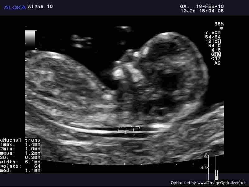
FIRST trimester screening is an important event for any pregnant woman. It is the first time they will see their baby and, weighing 45g and measuring 10cm from head to toe, some important features are already present in this 12-week-old foetus.
Ultrasound scans at this stage identify one important characteristic – the nuchal translucency (NL). This anatomical feature is a band of fluid behind the neck of a foetus [see Figure 1]. With 65-85% of babies with thickenings of the nuchal NT later diagnosed as having a trisomy disorder - chromosomal abnormality where there are three copies of one chromosome, instead of the normal two – this is an important indicator of trisomy risk.
Down’s Syndrome is a trisomy disorder, with three copies of chromosome 21. Characteristics of this syndrome include short stature, heart defects, learning difficulties and digestion problems. However, although there is a spectrum of these problems, many Down’s Syndrome sufferers will be unable to live independently.
The risk of a mother giving birth to a baby with Down’s Syndrome increases with the mother’s age: from 1 in 350 at the age of 35 to 1 in 100 for mothers aged 40. This risk is so well established, that many developed countries now routinely screen older mothers for the condition.
The current technique of using ultrasound to measure the NT is, as with all ultrasound techniques, user-dependant, meaning results depend on the skill of the clinician in positioning the probe and interpreting the image
Down’s Syndrome is diagnosed by an amniocentesis; taking a sample of fetal genetic material, but this carries its own not-insignificant risk. The risk of a miscarriage is roughly 1%, so referral to amniocentesis needs to be made using reliable and accurate determination of indicating factors.
The current technique of using ultrasound to measure the NT is, as with all ultrasound techniques, user-dependant, meaning results depend on the skill of the clinician in positioning the probe and interpreting the image. This can result in two very experienced sonographers getting very different NT values from the same patient. With such a risky and invasive procedure pending on these results, this level of subjectivity is unacceptable.
As Professor Kypros Nicolaides, a professor of fetal medicine at King’s College London, said recently: “Over the last 20-years, research has proven time and time again the NT test is the single most important marker of chromosomal abnormalities. Yet it still relies upon a human judgment, and accordingly there is often considerable variance between readings.”
This level of user dependency is found across all scans. Settings need to be adjusted for each patient, depending on a number of different factors. This time-consuming programming cannot be done in advance of the patient arriving.
Professor Nicolaides said: “When you first get an ultrasound machine from the manufacturer, you cannot just take it straight out of the box and start using it with patients. Unfortunately, each machine needs to be calibrated for different functions and for each individual patient.”
Over the last 20-years, research has proven time and time again the NT test is the single most important marker of chromosomal abnormalities. Yet it still relies upon a human judgment, and accordingly there is often considerable variance between readings
Additional issues with first trimester screening include the type of probe used. There are two main types currently used for pregnancy scanning; linear and convex probes. The linear probe provides good near field resolution, while the convex probe affords a more peripheral view. The two probes have come in and out of favour over the last couple of decades. The convex probe is used out of habit more commonly, but as doctors want more details about the fetal organs, linear probes are becoming increasingly more popular as they provide better resolution for first trimester screening. Changing habits and training in linear probe use is an important challenge for sonographers moving forward.
Working closely with Aloka, Professor Nicolaides has been developing a way to automatically generate this important measurement to minimise user dependency and associated errors.
The importance of accurate and reliable NT measurements was reflected in the establishment of the Nuchal Translucency Quality Review (NTQR) in 2002 to deal with the issues surrounding it1. The wider context of this screening must not be forgotten, since it has considerable emotional impact on the parents-to-be. In March 2011, the NTQR said ‘appropriate education, NT credentialed physicians and sonographers, adequate equipment and counseling resources are essential for practices offering NT measurement’. And The Fetal Medicine Foundation UK offers training on performing this 11-13 week screen to ensure mothers-to-be receive the best treatment2.
Automated NT needs to be formally peer reviewed and tested before it becomes accepted practice. However, from the results we have so far, it is clear we can already say this conclusively works and are now just waiting on verification
The widespread use of the scan, even in its current subjective form, is evidence of its power as a tool for obstetricians. In the last two years the test has become more widely available on the NHS; an indication of its clinical use in reducing amniocentesis referral rate. Professor Nicolaides said: “Automated NT needs to be formally peer reviewed and tested before it becomes accepted practice. However, from the results we have so far, it is clear we can already say this conclusively works and are now just waiting on verification.”
In fetal medicine, like all fields, current practices must be continually developed further. Over the next few years, these three improvements to first trimester screening will increase the accuracy and reliability of this scan; automated-Nuchal Translucency measurements, a linear probe, and pre-calibrated ultrasound machines. Being able to vastly improve the detection of chromosomal disease and reduce the human error factor and subjectivity in many of these tests is a significant development in fetal medicine.


