Artificial intelligence (AI) could be around twice as accurate as a biopsy at grading the aggressiveness of some sarcomas, according to new research from The Royal Marsden NHS Foundation Trust and The Institute of Cancer Research, London.
Results from the study, which have been published in The Lancet Oncology, suggest that a new AI algorithm could help tailor the treatment of some sarcoma patients more accurately and effectively than a biopsy, an invasive procedure which is currently standard practice.
The research also suggests that the technology could help clinicians diagnose subtypes of the rare disease, speeding up diagnosis.
And researchers believe the technique could be eventually applied to other cancer types, too, potentially benefitting thousands of patients every year.
Soft tissue sarcoma is a type of cancer that develops in the body’s connective tissues, including fat, muscles, nerves and blood, and lymph vessels.
Through this early research, we’ve developed an innovative AI tool using imaging data that could help us more accurately and quickly identify the type and grade of retroperitoneal sarcomas than current methods
It is a rare cancer, with around 4,295 people in England diagnosed each year and over 50 different types.
This study focused on retroperitoneal sarcoma, a soft tissue sarcoma which develops in the back of the abdomen and, due to its location and rarity, is currently hard to diagnose and treat.
Researchers used the CT scans of 170 Royal Marsden patients with the two-most-common forms of retroperitoneal sarcoma – leiomyosarcoma and liposarcoma – to create an AI algorithm, which was then tested on nearly 90 patients from centres across Europe and the US.
They used a technique called radiomics to analyse the CT scan data, which can extract information about the patient’s disease from medical images, including data which cannot be distinguished by the human eye.
The model accurately graded the risk – or how aggressive a tumour is likely to be – of 82% of the tumours analysed, while only 44% were correctly graded using a biopsy.
The model also accurately predicted the disease type of 84% of the sarcomas tested – meaning it can effectively differentiate between leiomyosarcoma and liposarcoma – compared with radiologists who were not able to diagnose 35% of the cases.
By giving clinicians a more-accurate and effective way of grading tumours, researchers hope this technology will improve the management of the disease and outcomes.
There is an urgent need to improve the diagnosis and treatment of patients with retroperitoneal sarcoma, who currently have poor outcomes
For example, as high-grade tumours can indicate aggressive disease, this tool could help ensure high-risk patients are identified and get amplified treatment, while low-risk patients could be spared unnecessary treatments, follow-up scans, and hospital visits.
It could also speed up diagnosis of the disease by supporting clinicians – who may never have previously seen a retroperitoneal sarcoma due to its rarity – in more accurately identifying the subtype.
The study was supported by funding from The Royal Marsden Cancer Charity, the National Institute for Health and Care Research (NIHR), the Wellcome Trust, and the EORTC Soft Tissue and Bone Sarcoma Group.
First author, Dr Amani Arthur, registrar at The Royal Marsden NHS Foundation Trust and Clinical Research Fellow at The Institute of Cancer Research, London, said: “There is an urgent need to improve the diagnosis and treatment of patients with retroperitoneal sarcoma, who currently have poor outcomes.
“The disease is very rare – clinicians may only see one or two cases in their career – which means diagnosis can be slow.
“This type of sarcoma is also difficult to treat as it can grow to large sizes and, due to the tumour’s location in the abdomen, involve complex surgery.
We are incredibly excited by the potential of this state-of-the-art technology, which could lead to patients having better outcomes through faster diagnosis and more-effectively-personalised treatment
“Through this early research, we’ve developed an innovative AI tool using imaging data that could help us more accurately and quickly identify the type and grade of retroperitoneal sarcomas than current methods.
“This could improve patient outcomes by helping to speed up diagnosis of the disease, and better tailor treatment by reliably identifying the risk of each patient’s disease.
“In the next phase of the study, we will test this model in clinic on patients with potential retroperitoneal sarcomas to see if it can accurately characterise their disease and measure the performance of the technology over time.”
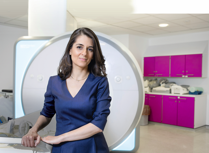
Study lead, Professor Christina Messiou, consultant radiologist at The Royal Marsden NHS Foundation Trust and Professor in Imaging for Personalised Oncology at The Institute of Cancer Research, London, added: “This is the largest and most-robust study to date that has successfully developed and tested an AI model aimed at improving the diagnosis and grading of retroperitoneal sarcoma using data from CT scans.
Our novel approach used features specific to this disease, but by refining the algorithm, this technology could one day improve the outcomes of thousands of patients each year
“Due to the rarity of the disease, this was a global effort and I am immensely proud and thankful to the team.
“We are incredibly excited by the potential of this state-of-the-art technology, which could lead to patients having better outcomes through faster diagnosis and more-effectively-personalised treatment.
“As patients with retroperitoneal sarcoma are routinely scanned with CT, we hope this tool will eventually be used globally, ensuring that not just specialist centres – who see sarcoma patients every day – can reliably identify and grade the disease.
“In the future, this approach may help characterise other types of cancer, not just retroperitoneal sarcoma.
“Our novel approach used features specific to this disease, but by refining the algorithm, this technology could one day improve the outcomes of thousands of patients each year.”
And Dr Paul Huang, team leader of the molecular and systems oncology team at The Institute of Cancer Research, London, said: “It is exciting to see AI – which is trained to spot incredibly-subtle signs that a cancer is aggressive from a simple CT scan – enable rapid diagnosis and classification of this rare type of sarcoma.
“Sarcomas represent a biologically-complex group of cancers – encompassing many distinct types – and telling them apart with the human eye poses a formidable challenge, particularly outside of specialist centres.
"This kind of technology has the potential to transform the lives of people with sarcoma – enabling personalised treatment plans tailored to the specific biology of their cancer. It is great to see such promising findings.”


