
Gamma Knife has become a recognised treatment for small to medium-sized tumours and vascular disorders that develop in the brain and skull base, including secondary metastasised tumours and arteriovenous malformations (AVMs), for example.
As a form of stereotactic radiosurgery, it presents a treatment option for complex cases where open surgery would carry significant risk.
Gamma Knife uses 192 precisely-focused beams of radiation which converge to produce a high dosage of radiation to a highly-focused target.
This means it is capable of treating complexly-located lesions without damaging the surrounding healthy brain tissue.
Gamma Knife has become a recognised treatment for small to medium-sized tumours and vascular disorders that develop in the brain and skull base
This year marks the 10-year anniversary of the Gamma Knife installation at the Queen Square Radiotherapy Centre (QSRC) within the University College London Hospital (UCLH); a centre of clinical excellence that operates within the National Hospital of Neuroscience and Neurology (NHNN).
A complex installation
 Providing stereotactic radiosurgery treatment options using Gamma Knife for complex lesions in the brain, this centre has been consistently listed as one of the top three facilities for neuroscience and neurology in the world.
However, the installation of the Gamma Knife unit was complex.
The process involved tonnes of equipment and live radio-active sources being fitted into the basement of UCLH – where the QSRC is located – which involved using cranes, creating a prefabricated breakout wall, and temporarily moving a steel staircase.
UK hospitals with medical ionising radiation facilities such as X-rays, nuclear scans, and radiotherapy units must meet the Care Quality Commission (CQC) regulations which aim to minimise risk of harm to patients and staff and set out the responsibilities of duty holders; to ensure what is needed for sufficient radiation protection and that the basic safety standards are met.
And, unlike linear accelerators (LINAC) such as teletherapy and radiotherapy units, which produce radiation electronically – only active when turned on – Gamma Knife machines house live Cobalt sources.
These are sourced from Canada, made using Cobalt 59 in a nuclear reactor which absorbs neutrons from the neutron flux in the reactor to become Cobalt 60.
A weighty challenge
Each Gamma Knife unit contains 192 pellets of Cobalt, each measuring 1mm x 1mm and these are transported in a nine-tonne cask.
After arriving on site, the sources are loaded into the Gamma Knife using a specifically-designed machine, which itself weighs three tonnes, and that removes the current pellets as well as transferring the new pellets into the Gamma Knife.
As the casks and loading machine themselves weigh tonnes, cranes are often needed for loading and unloading the sources, which are returned to Canada after the five-year half-life for disposal or re-use.
As the Cobalt 60 decays, it becomes weaker, and as the sources approach their five-year half-life, they have to be replaced.
The planning software for the machines automatically takes the source’s strength into account when planning treatments so clinical staff are aware of when it is approaching half life
The sources must be delivered in a flask source container, weighing 2-3 tonnes which then needs to have surface measurements taken on arrival to ensure it is sealed.
Although half as strong, the sources are still extremely strong after five years, up to 222 terabecquerels (1 TBq = 1 000 000 000 000 Bq- one becquerel being one unit of measurement of radioactivity). This means that there are 222TBq radioactive disintegrations per second.
Even so, after five years, treatment times become twice as long. Gamma Knife treatment length can vary, but for complex cases with multiple lesions can take several hours.
So, it is not logistically feasible for treatments to double in length every five years in terms of management.
The planning software for the machines automatically takes the source’s strength into account when planning treatments so clinical staff are aware of when it is approaching half life.
This means that hospitals looking to introduce a Gamma Knife must apply for a licence to house live radioactive sources on site.
Added protection
This is the same process for brachytherapy equipment and is in place to ensure conditions around security and safety are upheld by sites holding active sources.
The licence is granted by the Office for Administration of Radioactive Substances Advisory Committee (ARSAC) and includes requirements for tamperproof doors, lead-lined shielding for walls, etc.
As with many hospitals that deliver radiation, staff exposure is stringently monitored, with lead-lined PPE in place, just as it is with X-ray and LINAC technologies.
Radiation monitoring for staff in hospitals is an integral part of good practice and the core discipline for various roles, including RSOs, health physicists, medical physicists, radiologists, and nuclear medicine techs.
Hospitals must meet specialised infrastructural requirements to provide this treatment option, providing sufficient protection from radiation
This is because it is important to be acutely aware of exposure rates for staff.
But, as the Gamma Knife is self-shielding, radiation is only able to escape during treatment and at the QSRC treatment room, vinyl indications on the floor mark out the exposure in the area, in line with ARSAC conditions.
Because Gamma Knife requires housing this live radio-active source, hospitals must also meet specialised infrastructural requirements to provide this treatment option, providing sufficient protection from radiation.
The QSRC therefore uses specialised CAT4 doors, in line with the safety and security conditions, which not only insulate the radiation but are designed to withstand 40 minutes of power tools – protecting the cobalt source from theft.
Significant recent developments in Gamma Knife technology have included the release of the Icon model in 2015, which introduced the ability to offer frame-based and mask-based treatments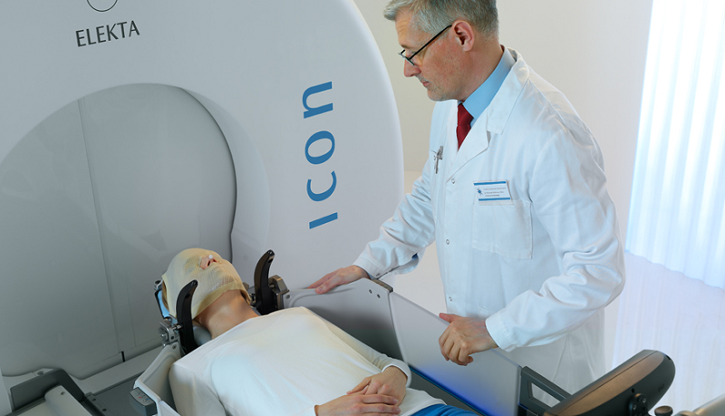
Proper planning
A multifaceted team comprised of architects, engineers, project managers, radiation safety advisors, radiologists, physicians, and chief technicians – who are going to be using the department – should be involved in the planning of a building with lead protection.
Generally, due to the small number of facilities that provide this service – which drives volume of work – it is highly desirable to plan the protection of a lead-lined room for the maximum-possible load.
This means, roughly speaking, the radiation protection of a general-purpose lead-lined room about 6 x 4 x 3 metres in size calls for a wall thickness in all directions – 2mm of lead.
And, while these treatment rooms require reinforced floors, prefabricated breakout walls, and lead lining, the Gamma Knife machines are self-shielding. So, there is only exposure risk when the Gamma Knife unit doors are open, during treatment.
Due to the small number of facilities that provide this service – which drives volume of work – it is highly desirable to plan the protection of a lead-lined room for the maximum-possible load
This means, as opposed to other radiation medical equipment, such as LINAC machines which require 2m-thick walls and specific power supplies, these rooms only require walls with a thin lead lining.
Such requirements are similar to that of a CT room, making Gamma Knife relatively flexible to implement and presenting an opportunity to convert old CT treatment rooms to house a Gamma Knife.
PIC ID="2"}
Ease of access
As the Gamma Knife requires lead shielding in the floors, walls, and ceilings, its installation was complex, especially being situated in the basement of a large London hospital (UCLH).
The Gamma Knife machine itself weighs approximately 20 tonnes, and the source changer used to load the sources weighs around 15 tonnes.
So, the installation required reinforced floors, closure of wards above, and delivery through a false wall in the treatment room.
Generally, the requirements for housing a Gamma Knife include ease of access, but the QSRC was a specific case, being ideally located in proximity to Queen Square imaging and Great Ormond Street Hospital.
Access to the centre presented as an issue, with two different types of cranes used and all buildings below evacuated during delivery.
And, with the equipment being so heavy, the biggest crane in Europe was used for the installation, which also involved a road closure through Westminster City Council.
Notably, transporting live radioactive sources is heavily regulated, but, moreover, the installation was during the 2021 London Olympics, which caused additional security concerns, further restricting access.
What enables this treatment to be as cutting edge and precise as it is, is the ability to plan where to target the Gamma Knife – a crucial element of the treatment process
However, the siting of a Gamma Knife centre is influenced by the associated facilities required, such as imaging.
A real advantage to operating a Gamma Knife facility is regular and efficient access to imaging.
Arguably the most-important part of this treatment is the planning – mapping out the lesions and determining where to target the Gamma Knife.
So, the QSRC is optimally located with the Queen Square Imaging Centre on the same premises.
And, although the technology is always evolving, the physical technology behind Gamma Knife has been used for decades.
What enables this treatment to be as cutting edge and precise as it is, is the ability to plan where to target the Gamma Knife – a crucial element of the treatment process.
The use of enhanced diagnostic imaging – for example on a 3T MRI – is to better identify the overall volume and target to treat more accurately and successfully.
Improved precision
And this is proving an exciting quality development introduced at QSRC this year.
As imaging possibilities advance and developments are made in MRI technology, this directly progresses the potential of Gamma Knife to become even more precise.
That being said, the Gamma Knife machines are designed with very few moving parts; the cobalt sources are housed within eight moveable sectors in the radiation unit that move forwards and backwards over predrilled holes in the tungsten that allow radiation to reach the patient during treatment.
As imaging possibilities advance and developments are made in MRI technology, this directly progresses the potential of Gamma Knife to become even more precise
Outside of that, the patient table is the only other moving part of the Gamma Knife, allowing for 99% uptime.
With a service contract and preventative maintenance carried out by the machine commissioning body, Elekta, components are replaced on a time-cycle basis and accuracy configuration is checked regularly.
This generally takes place once per quarter on an out-of-hours day when the clinic is empty.
The service and maintenance of the machines, therefore, tends to be straightforward, with the current capacity of the machines comfortably being able to treat 1,000 patients per year.
And, building on this, these machines are constantly being innovated with new developments being regularly introduced to improve ease of use.
New innovations
The most-significant recent developments have included the release of the Icon model in 2015, the new Vantage frame in 2018, and the new planning software released 2020.
The Icon model introduced the ability to offer both frame-based treatments and mask-based treatments, with the first addition of cones being used in the machine, as well as allowing for fractionated treatments.
And the release of the Vantage frame introduced a frame that was MRI compatible, allowing for clearer imaging for treatment planning.
The Vantage frame compliments the plan optimiser software, Lightning, that was released in 2020 by Elekta.
Also released at this time was a remote planning software which allows for remote planning – and thus made continuing treatments easier during COVID.
Not only does this eliminate the need for clinicians to physically be on site to plan a patient’s treatment, but gives clinicians the ability to choose the duration and conformality of treatments.
Post COVID, QSRC is seeing many more patients with multiple metastases, and this is where Gamma Knife, compared to other platforms, can offer particularly-effective treatment
They can therefore offer more-bespoke treatment sessions for their patients, which comes in particularly useful when treating more-frail patients, or those who are in the later stages of their cancer journey.
Providing Gamma Knife treatment entails operational direction and co-ordination.
Multiple imaging sessions per patient, specialised clinical training, long treatment periods, and exposure monitoring for staff can present implementation and treatment delivery challenges – especially considering the volume of patients seen with the cancer treatment backlog resulting from COVID.
QSRC has witnessed considerable growth in the volume of metastatic cases referred for Gamma Knife treatment.
And, post COVID, QSRC is seeing many more patients with multiple metastases, and this is where Gamma Knife, compared to other platforms, can offer particularly-effective treatment.


