A leading London oncology expert has praised the latest innovation in cancer treatment technology after a 38-year-old patient with a complex liver tumour beat the disease following a non-invasive procedure lasting just a few minutes.
Edward Leen, a professor of radiology at Imperial College London and a consultant in interventional radiology treatment at Hammersmith, Charing Cross, St Mary’s and The Princess Grace hospitals, claims the NanoKnife System could provide a much-needed solution for traditionally hard-to-treat cancers.
His comments come after a patient suffering from worsening and highly-involved liver cancer saw her tumour disappear after treatment with the device, which uses a series of electrical pulses delivered through needle electrodes to destroy cancer cells without harming surrounding tissue.
The technology, from AngioDynamics, can be used where standard radiofrequency ablation has failed, or in more complicated cases where tumours are located near to major blood vessels or in hard-to-treat areas of the body.
Depending on the size of the tumour, patients can undergo several sessions, each lasting just two minutes, and the treatment can be used in conjunction with chemotherapy to improve the chance of success.
Professor Leen explained: “NanoKnife is a safer technique compared to thermal-based ablative methods, with a faster recovery time, and it is much less painful. In addition, it has a synergistic effect when combined with chemotherapy, which makes it more effective and there is a lower risk of recurrence of the cancer.”
NanoKnife is a safer technique compared to thermal-based ablative methods, with a faster recovery time, and it is much less painful
Patients are admitted to hospital on the morning of the procedure and blood tests and cardiac tracing tests are performed. The patient is then given a general anaesthetic and their muscles are paralysed using a muscle relaxant. Two disposable NanKnife needle electrodes are then inserted through the skin and into the tumour using CT or ultrasound guidance.
Once the needles are in place, a series of 3,000-volt electrical pulses are delivered across the electrodes, completing the ablation process. The muscular paralysis is then reversed and the patient usually discharged the following day with a standard dose of paracetemol to control any pain or discomfort.
In the case of the liver cancer patient, previous treatment with the drug therapy, Mitotane, had failed, as had a right hepatectomy for rupture of the adrenocortial liver metastases and a repeat resection of the tumour.
With no other options available, she was offered ablation therapy and was put forward for treatment with the NanoKnife System.
The 38 year old patient had a complex liver tumour that other treatments had failed to eradicate
Professor Leen, who pioneered the treatment in the UK, said: “The main challenge was to remove a 6.5cm metastasis in the area of the liver that lay under the lung. It was also under the left ventricle of the heart, alongside the left hepatic vein and one of the main body veins, the inferior vena cava.
“There are a number of ways surgeons can ablate material and the first attempt at ablating this large metastasis was with conventional radiofrequency ablation (RFA), but due to the inadequacies of RFA in this situation, and a lack of precision with this technology, it was unfortunately not possible to completely remove all the tumour material.
“As a result, it was decided to try to use NanoKnife. Despite the location of the metastasis being so close to the heart and major blood vessels, the procedure was successful and uneventful and the patient made a full recovery and was discharged the following day.”
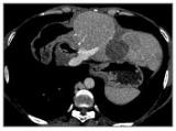
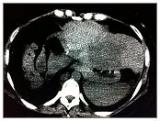
A CT scan performed six weeks after treatment showed a 50% reduction in tumour size
A subsequent CT scan performed six weeks after treatment showed a significant improvement in her condition, with an approximate 50% reduction in tumour size. Equally as important, all blood vessels that had been supplying the tumour had been destroyed; a critical step in ensuring the tumour cannot grow back again. Fifteen months after surgery, there has been no recurrence or return of the tumour.
The short-term outcome is very good, with 90% of our patients showing good control of the disease and a third showing shrinking of the tumour within four weeks of the procedure
Professor Leen said further trials were needed to test the long-term effects, but claimed it was a potential breakthrough in the treatment of complex cancer cases. He added: “This is a relatively novel procedure and as such there are no long-term efficacy results. However, the short-term outcome is very good, with 90% of our patients showing good control of the disease and a third showing shrinking of the tumour within four weeks of the procedure.”
However, there are some limitations to the system. For example, it cannot be used on patients with a pacemaker or a history of recent cardiac disease.